


Cost-effective method of adding 3D to X-Ray imaging
Conventional X-ray imaging equipment can now be upgraded to produce more advanced 3D images. Instrumentarium Imaging, a Finland-based specialist in X-ray imaging technology, is demonstrating new possibilities with its proprietary TACT technology. This technique is applicable in medical diagnosis as well as dental imaging applications.
"This approach is a more cost-effective way of producing such images without the need to make major investment in CT (Computed Tomography) equipment, for example," explains Timo Ihamäki, product manager at Instrumentarium Imaging. It will be possible to see TACT in action at RSNA'98."The TACT system offers a new way of looking at X-rays." He says that this software-based system offers not only greater accuracy, but also the ability to manipulate the X-ray images in different ways.
Images can be viewed from a variety of angles, rotated, or viewed in slices which is useful in biopsy procedures to provide more detailed information about the nature of underlying tissue. Such features are particularly important in medical diagnostics and treatment planning such as in mammography and sophisticated dental implant procedures.
Inherent accuracy
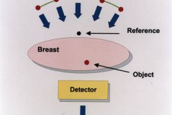
In addition, the fact that the TACT (Tuned-Aperture Computed Tomography) technology, that is widely patented, uses a reference point set in each X-ray image means that the accuracy of the three dimensional image comes from the image itself and not from the system mechanics. Typically, seven separate two dimensional X-ray images taken at different angles are used to create a 3D one. As each image is taken at 1/7th of normal radiation, no
dose increase is required.
One of the first instruments to exploit this technique is the Delta 16 TACT system which is based on the company's existing digital X-ray equipment used in mammography and biopsy. The company is planning to add this feature to its range of digital imaging systems. This will include full field digital mammography, the company's Delta DX system, as well as in Delta 16 for biopsy, its c-arm technology used during surgical procedures and its panoramic Orthopantomograph X-ray range used in dentistry.
Captions:
Figure 1: The third dimension can be added to existing medical X-ray imaging such as the Delta 16.
Figure 2: With a single reference point covering up to seven discrete X-ray images from different angles, three dimensional images can be built up.
For more information contact:
X-OGRAPH Imaging Systems Ltd.
Mr Neil Staff
X-Ograph House
Hampton Street, Tetbury
GLOUCESTERSHIRE GL8 8LD
ENGLAND
Tel.: +44 1 666 501 501
Fax.: +44 1 666 501 502
For medical imaging systems: Timo Ihamäki
For dental imaging applications:
Jyrki Ojanperä
Instrumentarium Corp.Imaging Division
Nahkelantie 160
PO Box 20
FIN 04301,Tuusula
Finland
Tel: + 358 9 258 851
Fax: + 358 9 275 7276
Web:http://www.instrumentarium.fi/imaging
Press release images
Cost-effective method of adding 3D to X-Ray imaging
Cost-effective method of adding 3D to X-Ray imaging

Feedback is not allowed / disabled for this press release.